Decellularization Studies in Regenerative Biology
How good would it be if we could produce artificial organs in the lab? So we could replace our worn and damaged organs, and we could live longer than we could possibly imagine before.
Realizing this dream may not be centuries away. There are many studies in the world to realize this. There are promising studies on this subject. For example, one of the most interesting of these studies is the mouse kidney that was "reproduced" and transplanted in the laboratory environment, which was the subject of the news a few years ago [1]. We say reproduced because decellularisation studies are used here, a new practice of regenerative biology. The decellularization process provides us with the organ skeleton required for artificial organ production.
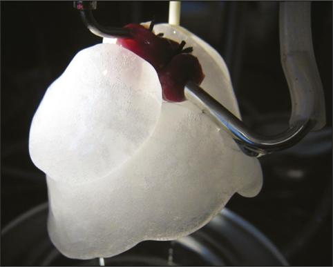
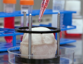
Figure 1: Decellularized natural heart skeleton awaiting heart cells [2]
So why do we need decellularisation instead of trying to build the organ skeleton from scratch? This is because, in today's conditions, it is extremely difficult to produce an organ skeleton from scratch instead of the natural one. The basis for this is that the cells can preserve their vitality at certain distances (1-3 mm) from the food source. In situations farther than these distances, it becomes impossible to produce thick tissues such as liver and muscle and to maintain their vitality. In a natural organ skeleton, the structures necessary for the cells to be fed are close to each cell to benefit from nutrients, so using the natural skeleton by filling it with new and functional cells is one of the effective methods for today's artificial organ technology. Organ decellularisation is the removal of cells from the organ using various chemicals containing detergents (chemicals used in cleaning and purification processes), resulting in an intercellular skeleton (for example, picture 1). New cells (especially stem cells) are placed in the natural 3D skeleton obtained as a result of this process, allowing new cells to form tissue. Since stem cells have the ability to transform into various cells, cells that have not yet undergone differentiation, cells that cannot function are produced from stem cells in the laboratory environment. Thanks to its ability to transform into the desired cell, stem cells have an important place in decellularization studies. [3]
An effective organ decellularisation is achieved as a result of the successful application of different chemical, biological or physical separation techniques. The methods used for successful organ decellularization vary depending on the tissue type. For fine tissue such as small intestine, pericardium, tissue is frozen and thawed. Then, the washing process is carried out using detergents that can be easily removed from the tissue. In thicker tissues such as the dermis, more biological and chemical substances are needed to remove cellular structures, and then more washing to remove the substance from the organ. For tissues and organs such as the pancreas and the brain that have a high fat content and do not have a certain shape, the result is achieved by adding lipid solvents and alcohols during the decellularization process [4].
Acids and bases, detergents, alcohols, hypertonic and hypotonic chemicals are used in chemical organ decellularization methods. Acids and bases used in decellularization are involved in the hydrolytic destruction of biomolecules and cytoplasmic components. For example, peracetic acid is an acid with antiseptic properties, which is frequently used in the process of decellularization of adipose tissues. Detergent, on the other hand, can be examined under two headings as ionic and nonionic detergents in the organ decellularization process. Non-ionic detergents are frequently used substances as they effectively cleanse the cells by not damaging the extracellular (extracellular) matrix structure in the tissue while decellularizing the organ. However, it does not affect interactions between protein-protein molecules while highly clearing the interactions between lipid-lipid and lipid-protein molecules. For example, Triton X-100 is the most commonly used nonionic detergent. It is mostly used for decellularisation of fine and delicate tissues (such as heart valves and blood vessels). Thanks to these properties, Triton X-100 can be called the most effective detergent for lipid elimination, but it also reduces the amount of glycosaminoglycan (GAG), one of the important components of the extracellular matrix, which is undesirable for decellularization [5].
Ionic detergents, on the other hand, help the cell breakdown process by dissolving the cell membrane and cause denaturation of proteins. SDS (sodium dodecyl sulfate) is an important ionic detergent and it can be said that it is more effective in decellularization process compared to Triton X-100, and as a result of these properties, it is used in decellularization of organs with higher cell density (such as lung and kidney). However, in addition to its effectiveness in decellularization, it reduces the amount of glycosaminoglycan in the extracellular matrix more than Triton X-100. This is an undesirable result that disrupts the mechanical balance of the skeleton. Also, because it is a more effective decellularizing detergent than Triton X-100, the incubation time is shorter than Triton X-100.
Alcohols, on the other hand, provide cell destruction by depriving cells of water (dehydration) in removing cellular structures. In lipid elimination (depilidization), the use of methanol and ethanol, the simplest alcohols, can be used in a certain amount, although the enzyme that provides lipid elimination is more effective than lipase because it causes protein precipitation and damage to the extracellular matrix. For this reason, methanol and chloroform mixed solutions are generally used for lipid elimination. [6]
Hypertonic and hypotonic solutions in decellularization help to remove cell debris from the organ. Hypotonic solutions disrupt the osmotic balance of the cell, leading to cell destruction. Some hypertonic solutions (hypertonic saline) allow DNA to be separated from proteins.
When it comes to biological methods, the process of removing cells from the organ as a result of the use of enzymes should come to mind. Trypsin, lipases and nucleases (RNases and DNAases) can be cited as enzymes primarily used in decellularization. Enzymes specifically remove cell debris or unwanted extracellular matrix debris in the tissue decellularisation process. In addition, enzyme residues that cannot be removed at the end of the procedures may cause a reverse immune effect and problems may occur while the tissue is refilled with cells [5].
Physical methods used in organ decellularization are temperature change, force, a special type of electroporation, NTRE (non thermal irreversible electroporation) and pressure. These methods generally lead to the destruction of cells and decellularization is performed by washing.
Scientists have performed many organ modeling using many decellularisations to realize their dreams of creating organs in the laboratory. Among the main ones are decellularisation in the lung, decellularisation of the heart, kidney and liver. In these organs, after decellularization, they are filled with appropriate cells and their functional again has been provided to a certain extent.
Decellularization in the Lung
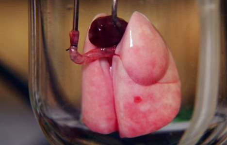
Figure 2: Recellular lung [7]
Lung diseases result in the death of hundreds of thousands of people every year in the world and are shown as the 4th cause of death in America. Pulmonary dysfunctions begin when it cannot regenerate sufficiently at the cellular level as a result of lung, lung cancer and other chronic diseases. Due to the low regenerative ability of the lung, the need for the lung to be produced by the organ decellularisation process has increased. Several different methods have been proposed for this. One of these methods is to decellularize the mouse lung with a detergent solution containing Triton X-100 and sodium deoxycholate, and then insert embryonic stem cells into the skeleton that will differentiate into the alveoli. As a result of skeletal (in vivo) cell cultivation called resellurization, embryonic stem cells differentiated into the alveole and microscopic vascularization (neovascularization) was observed. It is expected that such studies will help develop clinical approaches in the future.
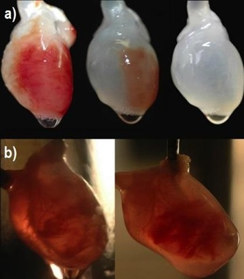
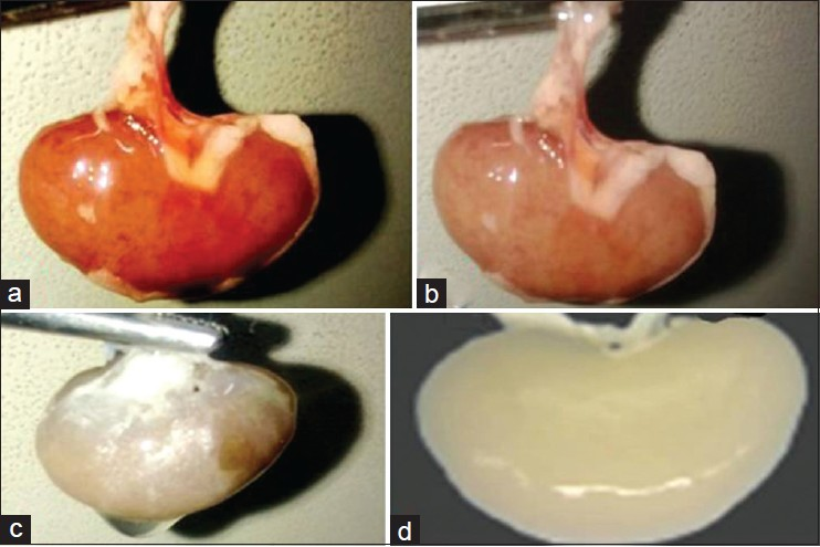
Figure 3a: A heart with step-by-step organ decellularisation
Figure 3b: Refilling the natural skeleton with cells
Decellularization and Recellularization of the Heart
Heart diseases are among leading causes of the death in the world. An artificial heart, which scientists have long dreamed of and produced with the patient's own tissues, may be the treatment that patients waiting for a heart transplant need. Producing a functional artificial heart can be achieved by applying a suitable 3D cardiac skeleton and the required cellular components to appropriate methods. Thus, to complete the decellularization process, appropriate detergents were applied to the mouse heart by coronary perfusion method, and as a result of the procedure, the extracellular matrix was preserved and cell-free vascular structures were obtained. Then, neonatal cardiac cells and mouse aortic endothelium cells were planted under physiological conditions suitable for organ development in order to regenerate cells into the obtained skeleton.First, he observed a contraction of the intermittent heart. Then, with the contraction of the myocardium, the pumping function of the heart was performed under appropriate electrical and other physiological stimuli.
Renal Decellularization
Picture 4: The decellularized kidney step by step [9]
Another decellularization application has been carried out in the kidneys. Advanced kidney failure can only be treated with kidney transplantation. Patients who cannot find a suitable kidney for transplantation may have to depend on dialysis device throughout their life. For these reasons, artificial kidney production studies are carried out, which can show the functional properties of the kidney. To date, primarily mouse, porcine and human kidneys have been decellularized, preserving the 3-dimensional skeletal structure of the kidney by perfusion of suitable detergents. Epithelial tissue cells and endothelium cells were then cultivated in the decellularized organ in a bioreactor under physiological conditions suitable for organ development. As a result of this process, an undeveloped form of urea was first detected in the organ in vivo. In addition, the intended urea production has been observed in vivo after the cellular kidney interacts with the mouse's circulation after transplantation into the mouse [10].
Liver Decellularization
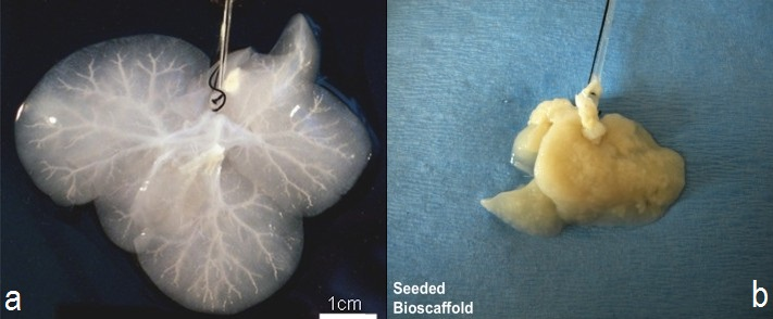
Figure 5a: A decellularized liver (decellularization)
Figure 5b: A liver with new cells planted (resellularization) [11]
A group of researchers at Wake Forest University has succeeded in decellularizing animal livers with a mild detergent with a mild detergent to produce artificial livers. As a result of decellularization, only the skeleton of the liver and other organs supporting the organ were left. Instead of liver cells, immature human liver cells and endothelial cells covering the blood vessels were planted into the skeleton through a large vessel. The liver was then placed in a bioreactor where nutrients and suitable biological conditions were provided for the organ. After 1 week, the researchers observed the formation of tissue that could indicate liver function [12].
As a result; As mentioned above with various examples, organ decellularisation is one of the main methods required for artificial organ production. Artificial organ technology is developing rapidly, with the production of different organs and tissues one by one day by day. Thanks to this technology, with the disappearance of the possibility of organ rejection since the organ will be filled with the patient's own stem cells, hopeful news can be given to patients waiting for organ transplantation and patients may not have to wait for the transplant.
Resources:
Laboratory production worked on kidney mice. Taken from http://www.bbc.com/turkce/haberler/2013/04/130415_bobrek-yapay.
MRS Bulletin Reviews Four Decades of Transformational Materials Developments. Taken from http://www.materials360online.com/newsDetails/42982.
Baptista, P. M., Siddiqui, M. M., Lozier, G., Rodriguez, S. R., Atala, A., & Soker, S. (2011). The use of whole organ decellularization for the generation of a vascularized liver organoid. Hepatology, 53 (2), 604-617.
Hrebikova, H., Diaz, D., & Mokry, J. (2013). Chemical decellularization: a promising approach for preparation of extracellular matrix. Biomed Pap Med Fac Univ Palacky Olomouc Czech Repub.
Crapo, P. M., Gilbert, T. W., & Badylak, S. F. (2011). An overview of tissue and whole organ decellularization processes. Biomaterials, 32 (12), 3233-3243.
Brown BN, Fruend JM, Li H, Rubin PJ, Reing JE, Jeffries EM, et al. Comparison of three methods for the derivation of a biologic scaffold composed of adipose tissue extracellular matrix. Tissue Eng Part C Methods. 2010
How To Build A Lung? Taken from http://speakingofresearch.com/2010/06/25/how-to-build-a-lung/.
University of Minnesota. "Beating Heart Created In Laboratory: Method May Revolutionize How Organ Tissues Are Developed." ScienceDaily. ScienceDaily, 14 January 2008.
Vishwakarma, S. K., Bhavani, P. G., Bardia, A., Abkari, A., Murthy, G. S. N., Venkateshwarulu, J., & Khan, A. (2014). Preparation of natural three-dimensional goat kidney scaffold for the development of bioartificial organ. Indian journal of nephrology, 24 (6), 372.
Sullivan, D. C., Mirmalek-Sani, S. H., Deegan, D. B., Baptista, P. M., Aboushwareb, T., Atala, A., & Yoo, J. J. (2012). Decellularization methods of porcine kidneys for whole organ engineering using a high-throughput system. Biomaterials, 33 (31), 7756-7764.
Researchers Make Miniature Human Livers In Lab. Taken from http://www.wakehealth.edu/WFIRM/.
Gilbert, T. W., Sellaro, T. L., & Badylak, S. F. (2006). Decellularization of tissues and organs. Biomaterials, 27 (19), 3675-3683.
Posted with STEMGeeks
Probably the can modelling these cell after decellularisation and produce a 3d printing file for mass production in the future
It's a good presentation. Thank you for sharing. Any idea why this post is muted?